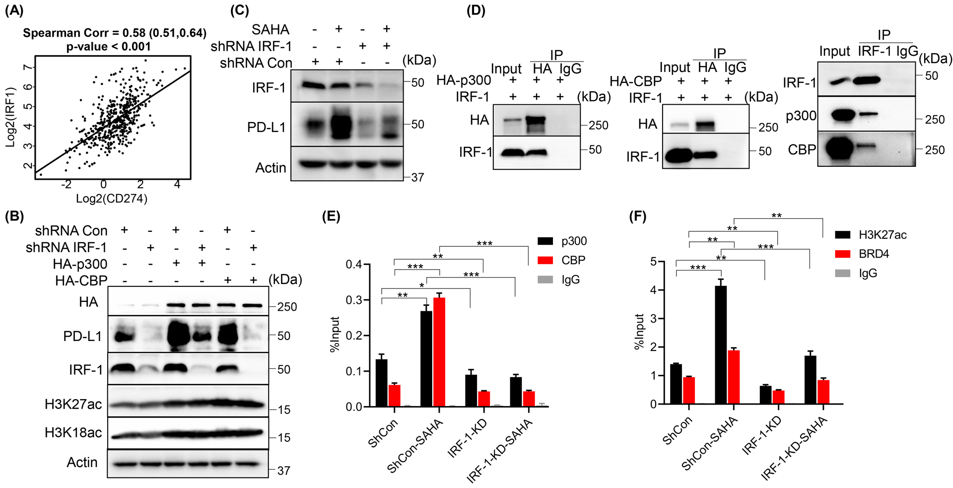
Figure 4. IRF-1 was involved in p300/CBP-induced PD-L1 expression in PCa cells.

(A)Scatter plot showed the correlation between (log2-transformed) expression of CD274 and IRF1 in prostate adenocarcinoma. IB analysis of WCL derived from DU145 cells infected with the indicated shRNA lentivirus followed by transfected with the indicated constructs (B) or 1 μM SAHA treatment (C). (D) IB of immunoprecipitate or WCL derived from DU145 cells transfected with the indicated constructs or not. (E, F) ChIP-qPCR analysis of p300, CBP, BRD4 and H3K27ac binding at CD274 promoter with primer P4 in DU145 cells infected with the indicated shRNA lentivirus followed by the treatment with 1 μM SAHA. All the statistical data were shown as mean values ± SD (n ≥ 3), *p < 0.05 **p<0.01 ***p<0.001.
