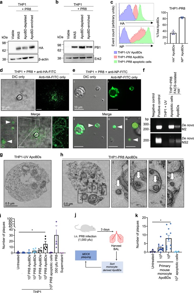
Fig. 2. Influenza proteins, mRNA, and virions are distributed into the ApoBDs of infected monocytes.
THP1 monocytes were infected with PR8 and ApoBDs were isolated 24 h p.i. via differential centrifugation for immunoblot analysis of HA a and PB1 b expression (representative of n = 2 independent experiments; WAS = Whole Apoptotic Sample). c THP1 monocytes were infected with PR8 (24 h p.i.) or subjected to UV irradiation (4 h), and ApoBDs and apoptotic cells were analysed for the presence of HA and NP by flow cytometry. d Live cell confocal microscopy was performed to monitor the distribution of HA on PR8-infected THP1 monocytes (24 h p.i.). e NP staining was monitored by confocal microscopy on PR8-infected THP1 monocytes (24 h p.i.). f PCR analysis of the 3ʹ spliced junction of IAV M2 and NS2 was performed on FACS-isolated HA+ apoptotic cells and ApoBDs. Transmission electron microscopy was performed on ApoBDs derived from UV-irradiated (4 h post UV) g or PR8-infected (24 h p.i.) h THP1 monocytes. i FACS-isolated ApoBDs from UV-treated or PR8-infected THP1 monocytes were subjected to viral plaquing (dots represent average of n = 2 biological repeats). j Schematic diagram of methodology used for plaque assay on primary monocyte ApoBDs. k Primary mouse monocyte ApoBDs were isolated via FACS from PR8-infected mice (1000 pfu, day 3 p.i.) and subjected to viral plaquing (dots represent average of n = 2 biological repeats). Unless otherwise specified, data shown is representative three independent experiments, *P < 0.05, unpaired Student’s two-tailed t-test.

