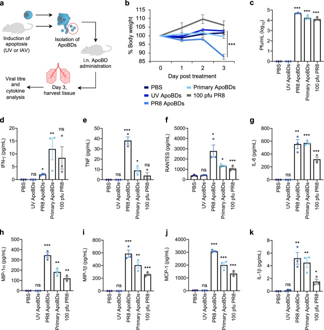
Fig. 4. Monocyte-derived ApoBDs can aid viral propagation in vivo.
a Schematic diagram of ApoBD isolation and in vivo administration. b Mice were treated with PBS, ApoBDs (UV-THP1, PR8-THP1, or primary mouse monocyte ApoBDs), or 100 pfu PR8, and body weight was monitored over 3 days post treatment. Lung tissue was collected on day 3 post treatment and lung viral titres were determined by MDCK plaquing c, and inflammatory cytokine production was determined by cytometric bead array d–k. Error bars represent SEM (n = 3 mice), ns = P > 0.05, *P < 0.05, **P < 0.01, ***P < 0.001, unpaired Student’s two-tailed t-test. Statistical significance of UV-ApoBD, PR8 ApoBD, Primary-ApoBD, and PR8 treatment was determined by comparison to the PBS only sample.

