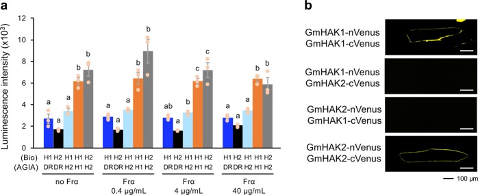
Fig. 3. Molecular interactions of GmHAKs.
a Luminescence intensities based on the AlphaScreen assay to assess the interactions between AGIA-conjugated proteins (AGIA) and biotinylated-proteins (Bio) in the presence and absence of Frα. All the individual data points are shown with the means and standard errors (n = 3). Means indicated by different small letters are significantly different, based on an ANOVA with post hoc Tukey’s HSD (P < 0.05). Recombinant proteins synthesized using a cell-free system are presented in Supplementary Fig. 12. DR, Escherichia coli dihydrofolate reductase serving as control; H1, GmHAK1; H2, GmHAK2. b Bimolecular fluorescence complementation (BiFC) analysis of GmHAK interactions. GmHAK1 or GmHAK2 fused to the N-terminal fragment of Venus (nVenus) and GmHAK1 or GmHAK2 fused to the C-terminal fragment of Venus (cVenus) were co-expressed in the epidermal cells of onions.

