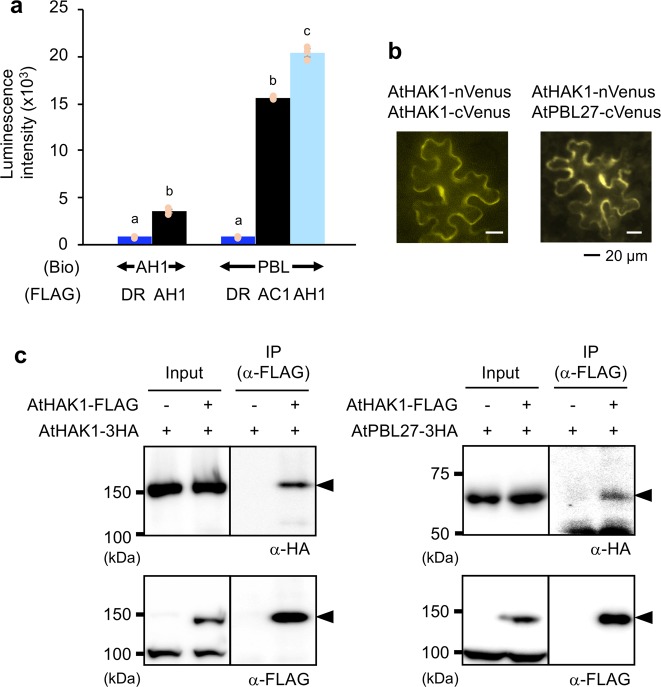
Fig. 7. Interactions of AtHAK1 and AtPBL27.
a Luminescence intensities based on the AlphaScreen assay to assess the interactions between biotinylated (Bio)-proteins for AtHAK1 (AH1) or AtPBL27 (PBL) and FLAG-conjugated proteins for Escherichia coli dihydrofolate reductase (DR) serving as control, AtCERK1 (AC1), and AtHAK1 proteins. Recombinant proteins synthesized using the cell-free system are presented in Supplementary Fig. 12. All the individual data points are shown with the means and standard errors (n = 3). Means indicated by different small letters are significantly different among the respective sets of data, based on a one-way ANOVA with post hoc Tukey’s HSD (P < 0.05). b AtHAK1 fused to the N-terminal fragment of Venus (nVenus) and AtHAK1 or AtPBL27 fused to the C-terminal fragment of Venus (cVenus) were co-expressed in Nicotiana benthamiana leaf cells. c FLAG-tagged AtHAK1 (AtHAK1-FLAG) and HA-tagged AtHAK1 (AtHAK1-3HA), AtHAK1-FLAG, AtHAK1-FLAG and HA-tagged AtPBL27 (AtPBL27-3HA) or AtPBL27-3HA were expressed in Nicotiana benthamiana leaf cells. Total proteins extracted from the leaves were immunoprecipitated using anti-FLAG-tag magnetic beads, subjected to SDS-PAGE, and probed with anti-HA or anti-FLAG antibody as a primary antibody. Arrowheads indicate the predicted, tagged AtHAK1 or AtPBL27 signals. Source data is presented in Supplementary Fig. 13.

