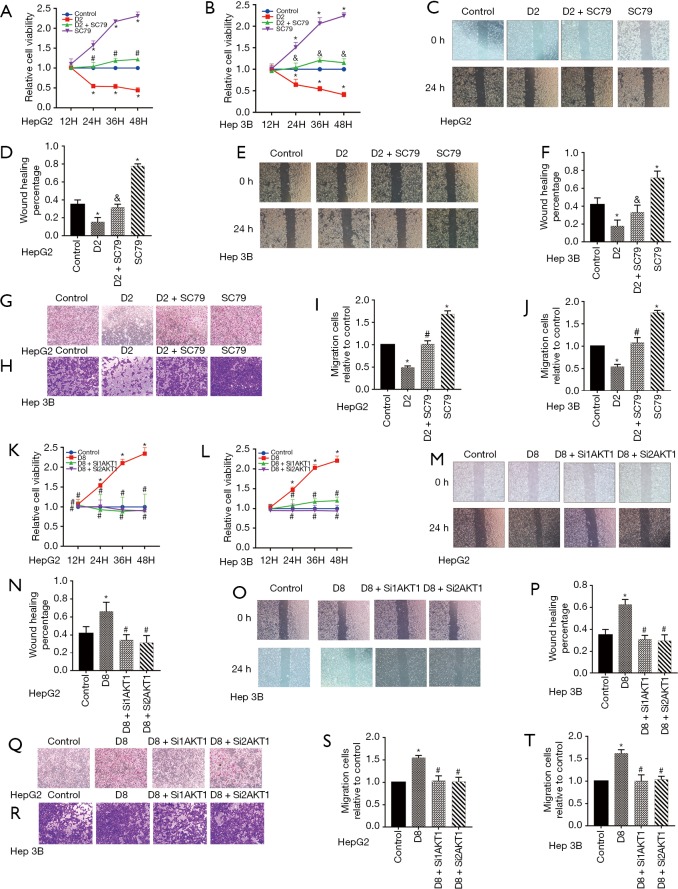
Figure 5.
Dezocine regulated HepG2 and Hep 3B cells viability and migration targeting Akt1/GSK 3β pathway. (A,B) Cell viability of HepG2 and Hep 3B cells in Medium containing dezocine of 2 µg/mL and Akt agonist SC79. (C,D,E,F) Cell migrate ability of HepG2 and Hep 3B cells treated by dezocine of 2 µg/mL and Akt agonist SC79 by wound healing assay (scale bar: 200 µm). (G,H,I,J) Cell migrate ability of HepG2 and Hep 3B cells treated by dezocine of 2 µg/mL and Akt agonist SC79 by transwell (cells were stained with crystal violet, scale bar: 200 µm). (K,L) Cell viability of HepG2 and Hep 3B cells treated with dezocine of 8 µg/mL and SiRNA. (M,N,O,P) Cell migrate ability of HepG2 and Hep 3B cells treated with dezocine of 8 µg/mL and SiRNA by wound healing assay (scale bar: 200 µm). (Q,R,S,T) Cell migrate ability of HepG2 and Hep 3B cells treated with dezocine of 8 µg/mL and SiRNA by transwell (cells were stained with crystal violet, scale bar: 200 µm). Data are represented as means ± SEM, n=3; *, P<0.05 vs. cells treated without dezocine; #, P>0.1 vs. cells treated without dezocine; &, P<0.05 vs. cells treated by SC79.

