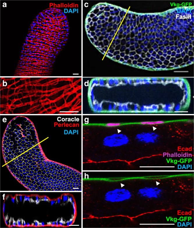
Fig. 1. Drosophila accessory gland recapitulates prostate epithelial micro- environment.

a, b alexa568-phalloidin (Phalloidin) staining reveals actin accumulation in the well-organised muscle sheet around the gland epithelium. b Higher magnification reveals actin bridges between the muscle fibres. This muscle sheet defines a stromal compartment in the gland. c–f Viking-GFP (GFP tagged Collagen IV, Vkg-GFP in (c, d) or Perlecan-RFP (Perlecan in (e, f)) expression reveals the presence of a basement membrane, and Fasciclin III (c, d) or Coracle (e, f) staining reveals the apico-lateral domain of the epithelial cells. Transversal optical section (along the yellow line in (c) and (e)) confirms deposition of Viking-GFP and Perlecan-RFP at the basal pole of the epithelial compartment. g, h Transversal optical section of the gland at high magnification. Viking-GFP expression reveals the presence of the basement membrane, E-Cadherin (Ecad) staining reveals the apico-lateral domain of one epithelial cell and alexa633- phalloidin (Phalloidin) staining reveals actin accumulation in the muscle fibres. Arrows indicate where the basement membrane encloses the muscle fibres. DAPI (blue) reveals nuclei in (a) and (b)–(h), mostly from binucleated epithelial cells (see g–h). Representative images in (a–d, g, h) from three or more experiments and in (e–f) from two. Scale bars: 50 μm in (a–f) and 10 μm in (g, h).
