
Fig. 5. Ras/MAPK and PI3K/AKT/TOR pathways are concomitantly activated in RasV12 expressing tumours.
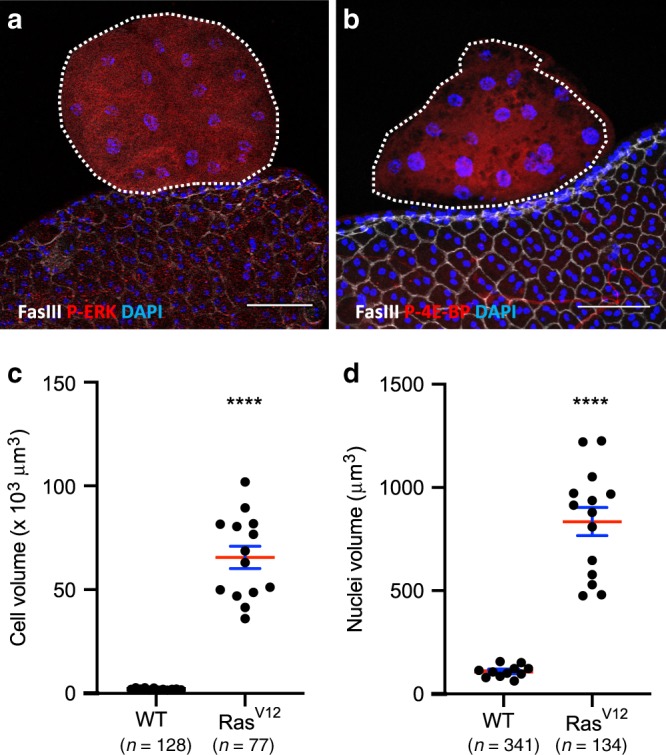
GFP-RasV12 expressing tumours are encircled by dotted lines. Fasciclin III (FasIII) staining reveals normal epithelial cells. a GFP-RasV12 expressing tumours display phosphorylation of ERK, a downstream target of the Ras/MAPK pathway. b GFP-RasV12 expressing tumours display phosphorylation of 4E-BP, a downstream target of the PI3//AKT/mTOR pathway. c, d Comparison of cell (c) and nuclei (d) volumes of tumour cells with wild-type cells (WT) (**** Unpaired t test; P < 0.0001) reveals strong hypertrophy of GFP-RasV12 expressing cells (RasV12). Data are represented as mean values±SEM (cell volume: n = 12 and n = 14 accessory glands Control and Rasv12 analysed, respectively, from seven experiments: nuclei volume: n = 11 and n = 14 accessory glands Control and Rasv12 analysed respectively, from seven experiments). Representative images in (a, b) from three or more experiments. DAPI (blue) reveals nuclei in (a, b). Scale bars: 50 μm.