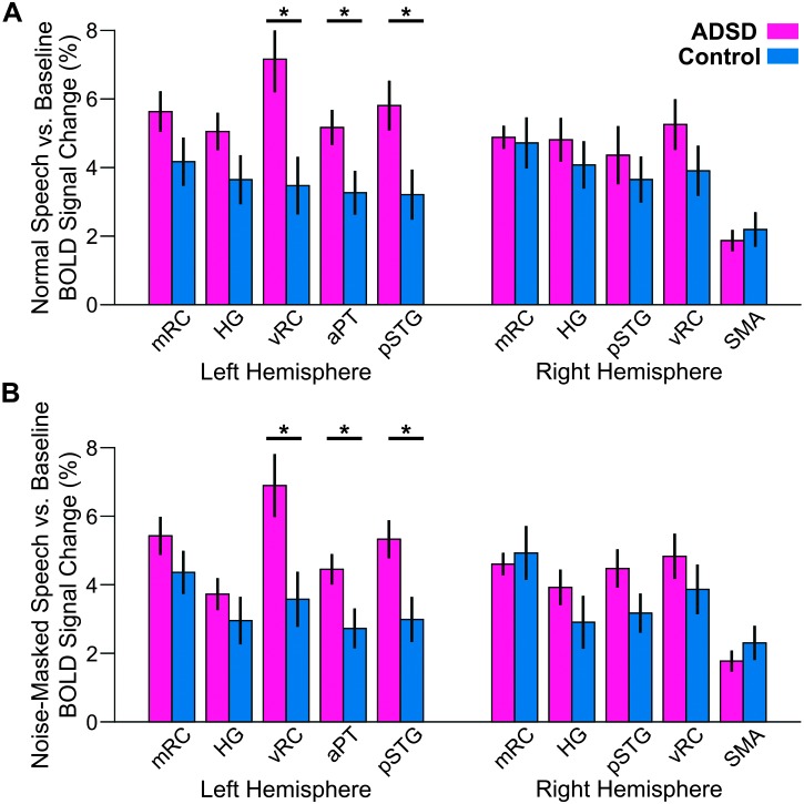
Figure 2.
Region of interest activity for the normal speaking (A) and noise-masked speaking (B) conditions contrasted with the baseline condition in the sentence production functional magnetic resonance imaging task for participants with ADSD (magenta) and control participants (blue). Significant group differences (p < .05, false discovery rate–corrected) are indicated by asterisks. See Figure 1 for region of interest definitions. Error bars correspond to standard error. BOLD = blood oxygen level dependent; ADSD = adductor spasmodic dysphonia; mRC = mid-Rolandic cortex; HG = Heschl's gyrus; vRC = ventral Rolandic cortex; aPT = anterior planum temporale; pSTG = posterior superior temporal gyrus; SMA = supplementary motor area.

