
Figure 3.
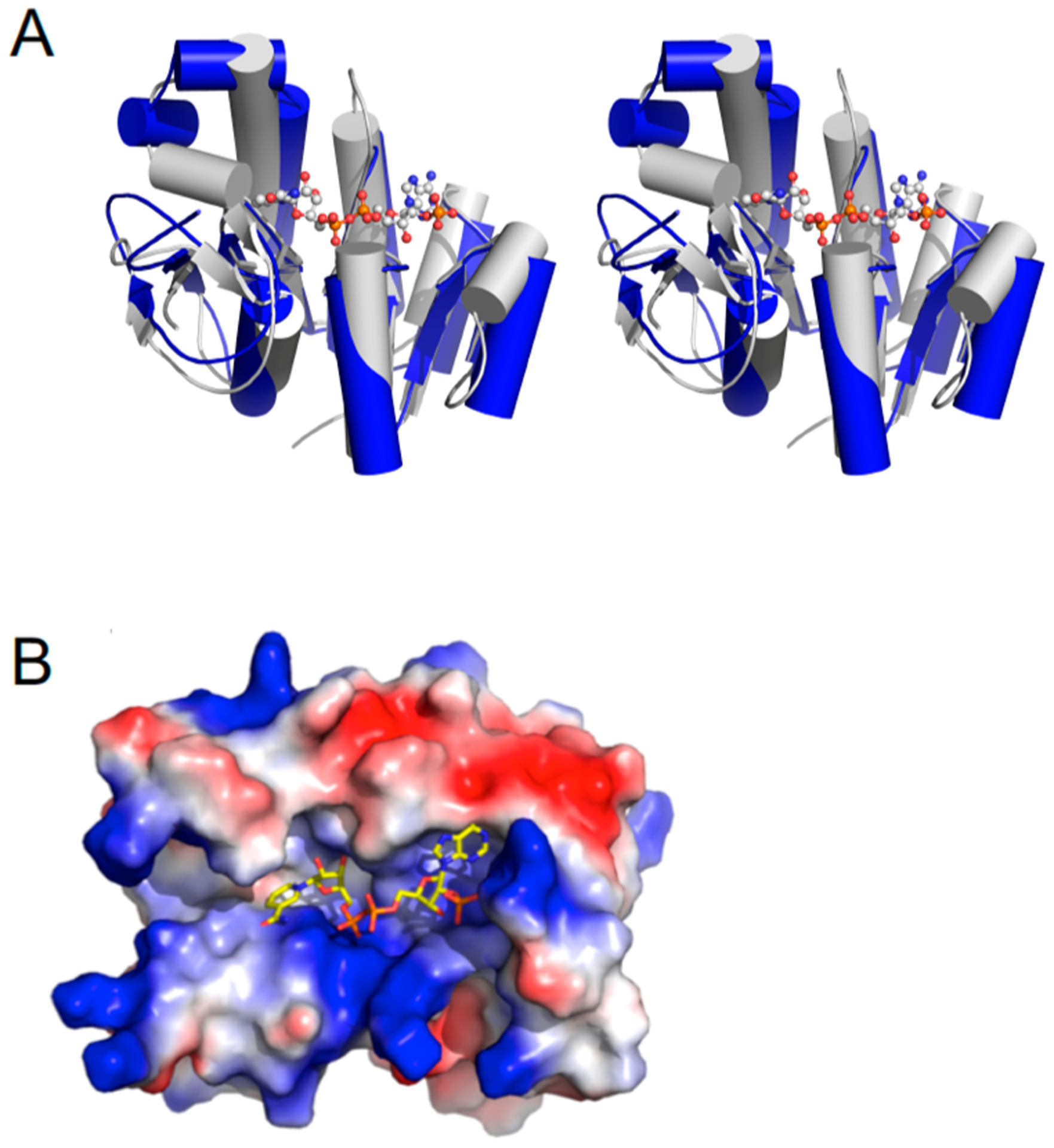
Structural alignment of ChuY with biliverdin β reductase (PDB entry 1HE3). (A) Wall-eyed stereoview showing a cartoon representation of ChuY (blue) aligned with biliverdin β-reductase (gray) by the Cα atoms that resulted in a rmsd of 3.06 Å2. (B) Same overlay showing only the cofactor from the biliverdin reductase model and the electrostatic surface representation of the ChuY model.