
Figure 4.
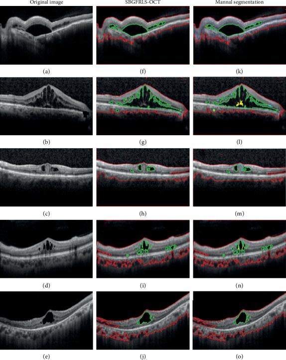
Comparison of DME segmentation performance between the SBGFRLS-OCT algorithm and manual method. (a–e) Original OCT images with DME pathology. (f–j) DME segmentation result by SBGFRLS-OCT algorithm. (k–o) Manual segmentation result by five different clinicians. Red lines were used to label the retinal region (ROI). Green lines were used to label DME regions.