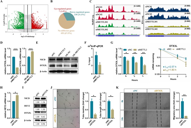
Fig. 4.
METTL3 activates Notch pathway via reducing the expression of DTX3L in endothelial cells. a Volcano map showing the m6A enrichment peaks in METTL3 deficient endothelial cells compared to control. Significantly increased and decreased peaks (fold change > 2, p-value < 0.001) are highlighted in red and green, respectively (b) Pie chart displaying the transcription level of genes with reduced m6A modification. c Integrative Genomics Viewer (IGV) tracks displaying MeRIP-seq and RNA-seq read distribution in DTX3L mRNA of control and METTL3 deficient cells. d qRT-PCR and (e) western blot analysis of the expression level of target genes. f m6A-IP-qPCR analysis of m6A enrichment on DTX3L mRNA in control and METTL3 deficient cells. g The mRNA half-life of DTX3L transcript in control and METTL3 depletion endothelial cells. h qRT-PCR and (i) Western blot analysis of indicated genes in control and DTX3L deficient endothelial cells. j Effects of DTX3L on tube formation and (k) migration of endothelial cells. Data are shown as mean ± SEM of three independent experiments. P values were calculated using Student’s t-test. *, P < 0.05; **, P < 0.01; ***, P < 0.001. P < 0.001

