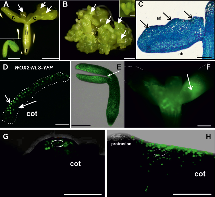
Fig. 3.
Development and symplasmic communication of cultured 35S:BBM IZE explants. (A) Explant at day 10. Embryos appear along the length of the cotyledons (arrows; c, cotyledon; h, hypocotyl; r, root). Inset, explant morphology at the beginning of the culture. (B) Groups of somatic embryos (arrows) after 28 d of culture; secondary somatic embryos form on the primary embryos (inset). (C) Explant after 4 d of culture; arrows indicate protrusions on the adaxial (ad) side of the cotyledons (ab, abaxial). (D) Expression of WOX2:NLS-YFP in a few layers of protodermal and subprotodermal cells in the adaxial side of a 3-day-old explant cotyledon (cot). The dotted white line demarcates the area engaged in SE. (E) One-day-old explant, 30 min after applying HPTS; the explant is still a single symplasmic domain. The white arrow indicates the site of fluorochrome application. (F) Seven-day-old explant, 30 min after HPTS application on the cotyledon node. The white arrow indicates the site of fluorochrome application. The fluorochrome moved through the explant, with the exception of the distal parts of cotyledons. (G) Six-day-old explant after CMNB uncaging in an embryogenic protrusion on the adaxial side of cotyledon (cot). The dotted white ellipse marks the uncaging area. (H) Six-day-old explant after CMNB uncaging in a non-protruding (non-embryogenic) region of the explant. The dotted white ellipse marks the uncaging area. Scale bars: (A, A inset, B, B inset) 500 µm; (C, E, F) 100 µm; (D, G, H) 50 µm.

