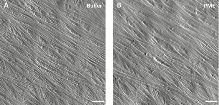
Fig. 7.
PME treatment reduces cellulose microfibril resolution by AFM imaging. (A) Peak force error image of an onion epidermal wall surface in HEPES buffer. Scale bar=20 nm. Distinct cellulose microfibrils and bundles are well resolved by AFM. (B) The same area scanned after PME treatment. The resolution of cellulose microfibrils is reduced while fibrils in the underlying lamella were obscured. Similar results were observed with six biological replicates.

