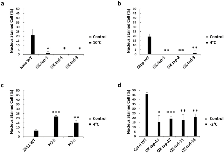
Fig. 4.
Determination of propidium iodide (PI) staining of nuclei in epidermal tissues of rice and Arabidopsis leaves using laser confocal microscopy. Nuclear staining indicates plasma membrane damage. (a) Percentage of cells with stained nuclei in the wild-type (WT) indica accession Kasalath (Kasa) and in three OsUGT90A1-overexpression (OX) transgenic lines grown under control conditions (28/25 °C day/night, 12-h photoperiod) or after chilling treatment at 10 °C for 2 d. (b) Percentage of cells with stained nuclei in the WT japonica accession Nipponbare (Nipp) and in three OsUGT90A1-OX transgenic lines grown under control conditions or after chilling treatment at 4 °C for 4 d. (c) Percentage of cells with stained nuclei in the WT japonica accession Zhong Hua 11 (ZH11) and in two OsUGT90A1-knockout (KO) lines grown under control conditions or after chilling treatment at 4 °C for 4 d. (d) Percentage of cells with stained nuclei in WT Arabidopsis Col-0 and in four transgenic lines overexpressing OsUGT90A1 (OX) grown under control conditions (22/20 °C, 16/8 h day/night) or after freezing treatment at –2 °C for 1.5 h. Ind- and Jap- indicate that the indica or japonica allele of OsUGT90A1 was overexpressed, respectively. Significant differences between the WT and the transgenic lines were determined using two-tailed Student’s t-tests: *P<0.05; **P<0.01; ***P<0.001. Note that almost no staining was observed under control conditions.

