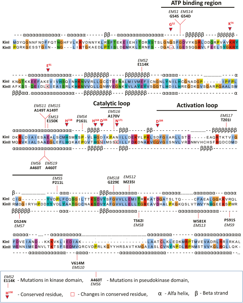
Fig. 5.
Primary and secondary structures of WTK1 kinase and pseudokinase domains alongside positions of knockout EMS mutations in yr15, yrG303, and yrH52 susceptible mutants. The diagram of WTK1 domain architecture highlights eight key conserved residues in the kinase domain (with numbers that correspond to their positions in cAPK (Hanks et al., 1988)) and the absence of five of them in the pseudokinase domain. Vertical lines indicate EMS mutations that block resistance. KinI, kinase domain; KinII, pseudokinase domain. (This figure is available in color at JXB online.)

