
Figure 3.
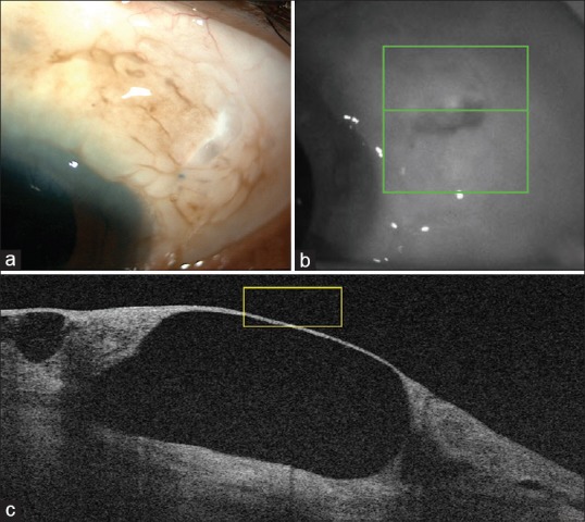
(a): Slit-lamp photograph of the left eye showing cystic bleb in the superotemporal quadrant. (b): Demonstration of the level of cross-section of optical coherence tomography. (c): Anterior segment optical coherence tomography through the bleb showing thin walled cyst