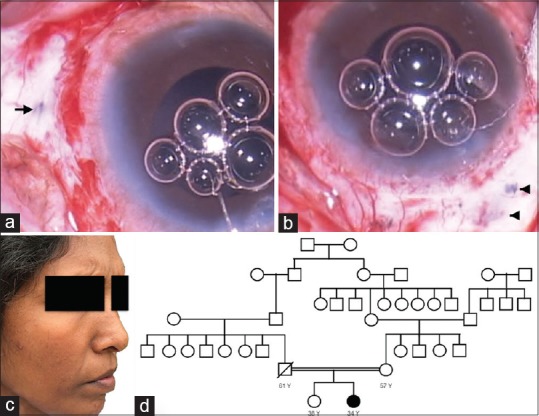
Figure 4.

(a): Linear area of thinning in the sclera (arrow) at 3 o' clock parallel to the limbus corresponding to sclerotomy. (b): Area of scleral thinning in the superonasal quadrant (arrow head). (c): Face photograph showing flat cheek. (d): Analysis of pedigree chart shows common ancestral origin of subject's parents. Pedigree chart is incomplete as she could not recollect beyond her grandparents
