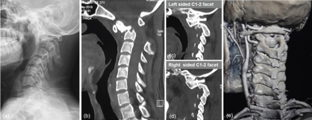
Figure 1.

(a) Lateral radiograph of cervical spine with anterior translation of C1 over C2 and fractured and anteriorly displaced dens.(b) Sagittal view computed tomography (CT) showing Type 2 odontoid fracture with anterior displacement and angulation. (c and d) Sagittal cuts through the facet joints showing locked right and left facet joints. (e) CT angiogram done to see anomalous vertebral artery anatomy showing exposed superior articular facets of C2.
