Figure 2.
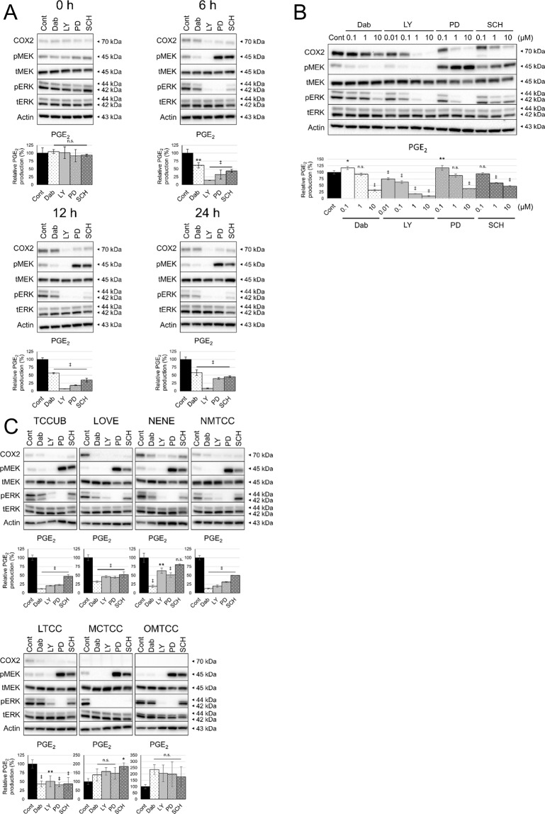
Effect of ERK MAPK inhibition on COX2 expression and PGE2 production. Protein levels in whole cell lysates were detected by Western blotting with Actin as loading control. Amount of PGE2 in culture medium was measured by enzyme-linked immunosorbent assay and normalised to cell number. Bar graph represents % control of PGE2 production. (A) cUC cells (Sora) were treated with vehicle (dimethyl sulfoxide; Cont) and inhibitors of BRAF (Dabrafenib; Dab), pan-RAF (LY3009120; LY), MEK (PD0325901; PD) and ERK (SCH772984; SCH) at 1 μM for indicated time. (B) cUC cells (Sora) were treated with vehicle (Cont), Dabrafenib (Dab), LY3009120 (LY), PD0325901 (PD) and SCH772984 (SCH) for 12 h at indicated dose. (C) cUC cell lines (TCCUB, Love, Nene, NMTCC, LTCC, MCTCC and OMTCC) were treated with vehicle (Cont), Dabrafenib (Dab) at 10 μM, LY3009120 (LY) at 1 μM, PD0325901 (PD) at 10 μM and SCH772984 (SCH) at 10 μM for 12 h. Data are presented as mean ± SD of three experiments. *Indicates p < 0.05, **p < 0.01, ‡p < 0.001 compared to vehicle control (Dunnett’s test).

