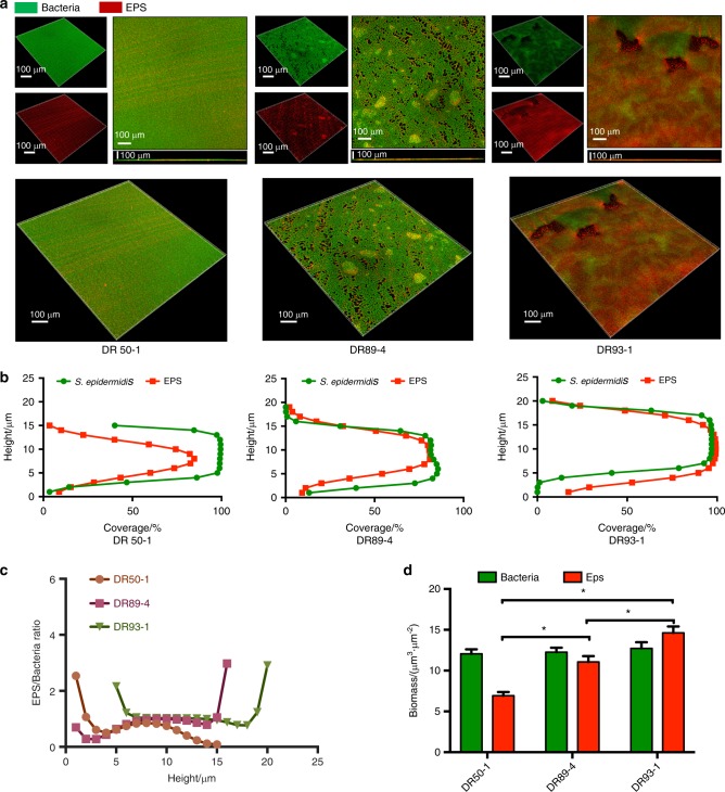
Fig. 5.
Biofilm architecture and EPS distribution of S. epidermidis strains observed by confocal microscopy. a Double-labeling of 24 h S. epidermidis biofilms. Green, bacteria (SYTO 9); red, EPS (Alexa Fluor 647). The three-dimensional reconstruction of the biofilms and the quantification of bacteria/EPS biomass were both performed with IMARIS 7.0.0. b The distributions of EPS and bacteria at different heights. c Quantification of bacteria/EPS biomass was performed with IMARIS 7.0.0. Results are the average of five randomly selected positions of each sample and are presented as mean ± standard deviation. P < 0.05. d The ratio of EPS to bacteria at different heights was quantified with IMARIS 7.0.0. Results are the average of five randomly selected positions of each sample and are presented as mean ± standard deviation. EPS, Extracellular Polymeric Substances

