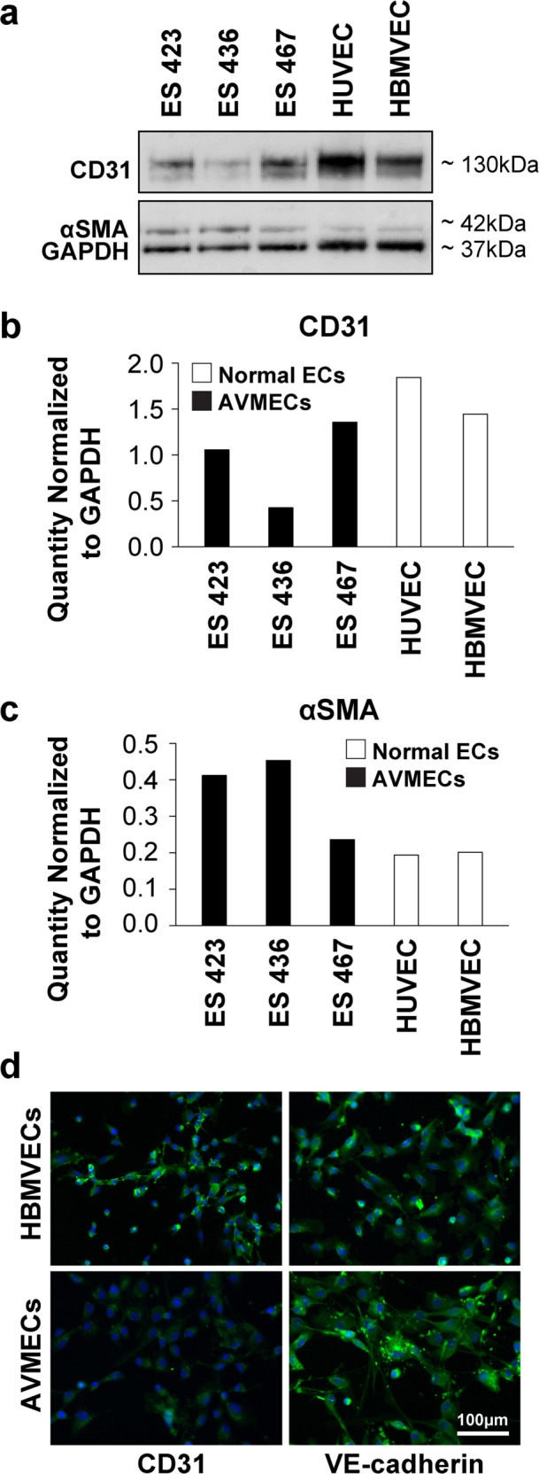
Fig. 1. Characterization of patient-derived AVMECs.

Primary endothelial cell lines were isolated from the human AVM tissue of three separate patients. The “purity” of primary patient-derived AVMECs was tested by Western blot (a) analysis using CD31 as an endothelial marker and αSMA as a mesenchymal marker. AVMECs express levels of CD31 (b) and αSMA (c) that are comparable with those of commercially available normal vascular endothelial cell lines. AVMEC endothelial identity was further confirmed by immunofluorescence that identified positive expression of endothelial lineage markers (CD31 and VE-cadherin). CD31 was also analyzed by Western blot and immunofluorescence, and VE-cadherin was analyzed by immunofluorescence (d) (CD31 and VE-cadherin = green, DAPI (nucleus) = blue). Original magnification ×200. Interestingly, VE-cadherin expression in AVMECs is not observed at the junctions, which is a pattern often noted in endothelial to mesenchymal transition.
