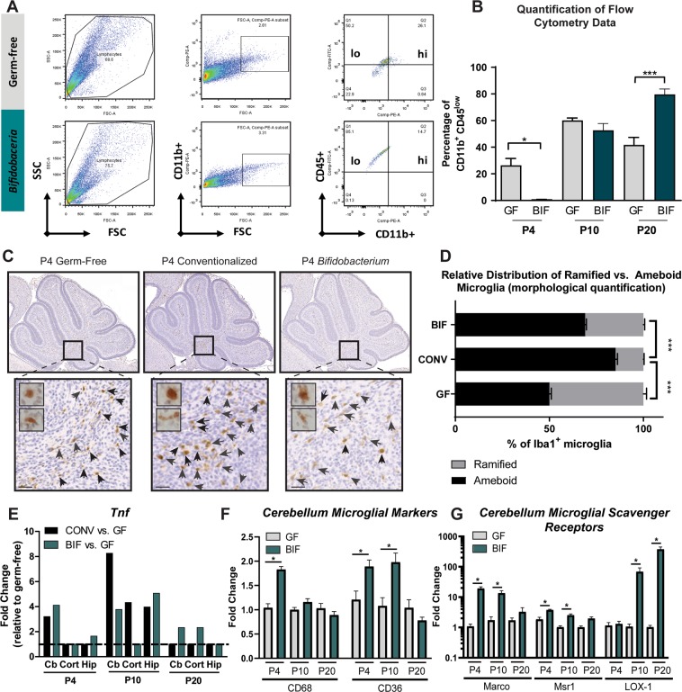
Figure 3.
Colonization with conventional microbiota and bifidobacteria facilitates early microglial reactivity and mid-late microglial ramification (a) Representative image of gating strategy for flow cytometric analysis. (b) Results plotted as percentage of CD11b+ CD45low microglia/total microglia at each timepoint. Ramified microglia possess the phenotype CD11b+,CD45low. At P4, GF mice have an increased % of microglia expressing markers of ramified microglia, relative to BIF mice. (One-Way ANOVA; *p<0.05, **p<0.01, *** p<0.001; n = 3 male animals/group/timepoint) (c) Microglia were labeled using an anti-IBA1 (Ionized calcium binding adaptor molecule 1) antibody. Scale = 50 μm. Representative images of staining in cerebellar white matter of P4 pups in each group, demonstrating fewer ameboid microglia in P4 germ-free mice (d) Morphological classification and quantification of ramified vs. ameboid microglia in P4 mice (**** p<0.0001, n = 3 mice/group, with 3 fields per mouse). (e) Expression of Tnf gene from whole tissue homogenate in each region (Cb = Cerebellum, Cort = frontal cortex, Hipp = Hippocampus) (data generated from PCR array data, therefore statistical significance and error bars are not shown for these pooled samples; n = 5 male mice/group/timepoint/brain region) (f) qPCR analysis of GF and BIF mouse cerebellum for markers of phagocytic microglia. (One-Way ANOVA; *p<0.05; n = 8 male animals/group/timepoint) (g) qPCR analysis of GF and BIF mouse cerebellum for scavenger receptors, which are required for proper phagocytosis. (One-Way ANOVA; *p<0.05; n = 8 male animals/group/timepoint).

