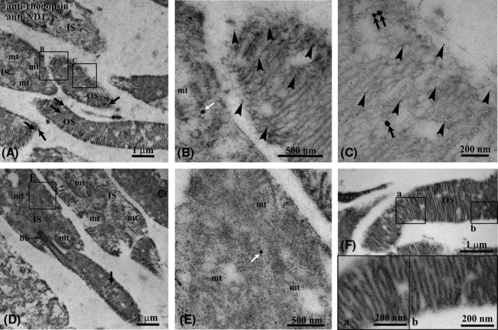
Figure 1.

Immunogold transmission electron microscopy experiment on bovine retina. A, Retinal section showing inner (IS) and outer (OS) photoreceptor segments. B and C, Enlargements corresponding to the squared areas B and C in panel (A), to show details of an OS. Largest gold particles (25 nm width) reveal Ab against ND1 in OS (black arrows) and in IS (white arrows). The smallest gold particle (5 nm width, arrowheads) reveals Ab against Rhodopsin that, as expected, is limited to the OS. The two Abs colocalize in OS. D, Additional retinal section showing an IS with a gold particle 25 nm width (white arrow) indicating the localization of ND1 in a mitochondrion. E, Enlargement corresponding to the squared area E in panel (D). F, OS of a negative control, in which the preimmune serum was applied instead of the specific primary Ab. No gold particles are visible. bb, cilium basal body; mt, mitochondrion; a, b: enlargements of the squared areas in panel (F)
