
Figure 2.
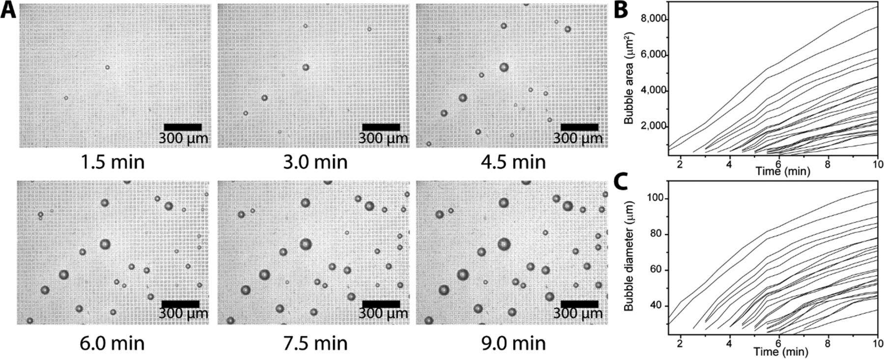
Kinetics of microbubbling. (A) Microscope images of the microbubbles growing on a section of the microchip (scale bars: 300 μm). About 25,000 Neutravidin functionalized platinum nanoparticles were incubated with biotinylated bovine serum albumin (bBSA) functionalized paramagnetic beads and loaded onto the microbubbling chip via external magnetic field. Time 0 was set as the point that the magnetic field was applied. Measurements of the microbubble (B) areas and (C) diameters as a function of time. Each trace represents the growth of one individual microbubble.