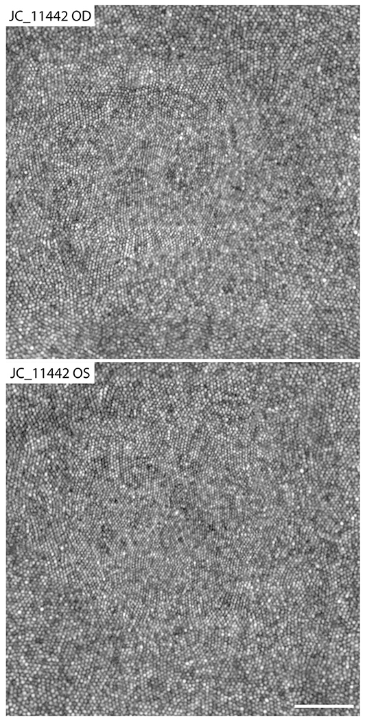
Figure 6.
Adaptive optics scanning light ophthalmoscopy images of the foveal cone mosaic for participant JC_11442. The right eye was determined to have a fragmented foveal avascular zone (FAZ) and shows a peak cone density of 241 286 cones/mm2. The left eye was classified as having a normal FAZ and showed a peak cone density of 247 710 cones/mm2. These peak cone density values are consistent with normal values from previous studies.39,45 Scale bar = 50 μm.

