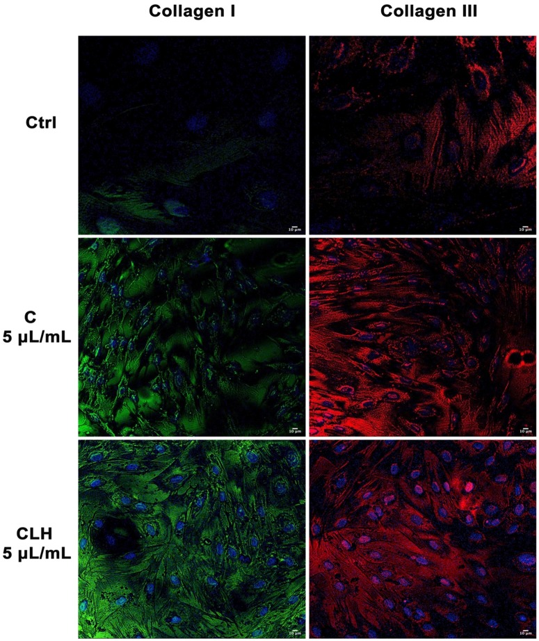
Figure 7.
Analysis of collagen deposition during wound healing. Immunohistochemical analysis of the expression of Collagen type I and type III was assessed in fibroblasts after scratch and treatment with different concentrations of single waste extract (C, L, H) or pooled waste extracts (CLH, CL, CH, LH) for 72 hours. Control cells were cultured in basic growing medium. Nuclei are labelled with 4,6-diamidino-2-phenylindole (DAPI, blue). Scale bars: 10 µm. The figures are representative of different independent experiments. For each differentiation marker, fields with the highest yield of positively stained cells are shown.

