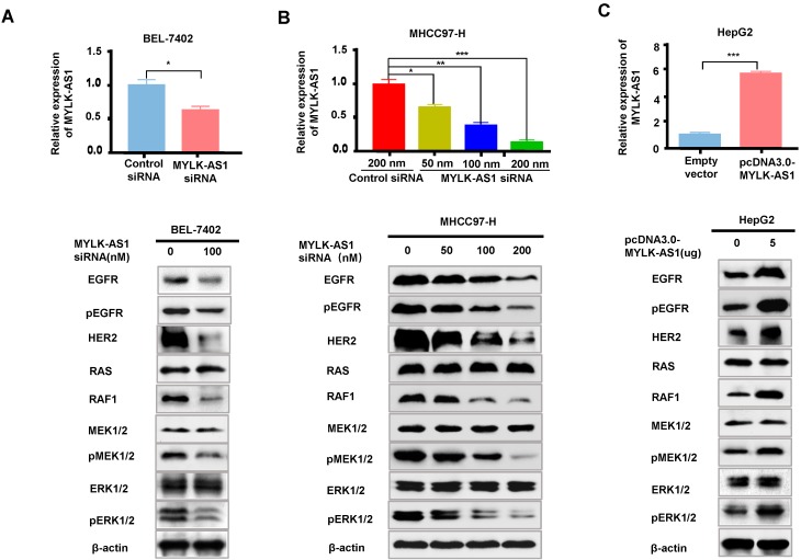
Figure 4.
MYLK-AS1 activates EGFR/HER2-ERK signaling pathway in HCC. (A) BEL-7402 cells were transfected with MYLK-AS1 siRNAs (100 nM) or control siRNA (100 nM). The MYLK-AS1 knockdown effect was detected by RT-qPCR. Western blot was performed to determine the expression of EGFR/HER2-ERK signaling pathway-related genes as indicated. β-actin was used as a loading control. (B) MYLK-AS1 siRNAs (50 nM, 100 nM and 200 nM) or control siRNA (200 nM) were transfected into MHCC97-H cells. The MYLK-AS1 overexpression effect was measured by RT-qPCR. Western blot was performed as in (A). (C) HepG2 cells were transfected with MYLK-AS1 (5 μg) or empty vector. The MYLK-AS1 overexpression effect was measured by RT-qPCR. Western blot was performed as in (A). All experiments were conducted three times independently and representative immunoblot results were shown. Data were presented as the mean ± SD (*P < 0.05, **P < 0.01).

