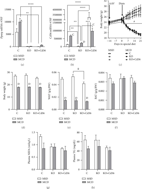
Figure 1.

Effect of MCD diet on body composition, plasma lipids, and ALT levels of PpargΔHep mice and PpargΔHep mice with overexpression of hepatocyte CD36. Hepatic expression of (a) Pparg and (b) Cd36. Gene expression is represented as an absolute copy number normalized by the normalization factor (NF). (c) Changes in body weight induced by MSD and MCD diets. (d) Body weight at sac. (e) Relative white adipose tissue (WAT) weight. The weight of WAT is the sum of urogenital and subcutaneous adipose tissue weights. (f) Relative brown adipose tissue (BAT) weight. Plasma (g) NEFA and (h) TG levels. Values are represented as the mean ± standard error of the mean. Letters or # represents significant differences between MSD and MCD within the group. Asterisks indicate significant differences between groups within the same diet. ∗,A,#p < 0.05; ∗∗,Bp < 0.01; ∗∗∗,Cp < 0.001; ∗∗∗∗,Dp < 0.0001. Control mice (C, circles); PpargΔHep mice (KO, squares); PpargΔHep mice with hepatocyte CD36 overexpression (KO+Cd36, triangles). MSD diet: open columns, open symbols; MCD diet: close columns, close symbols. n = 3‐7 mice/group.
