
Fig. 4.
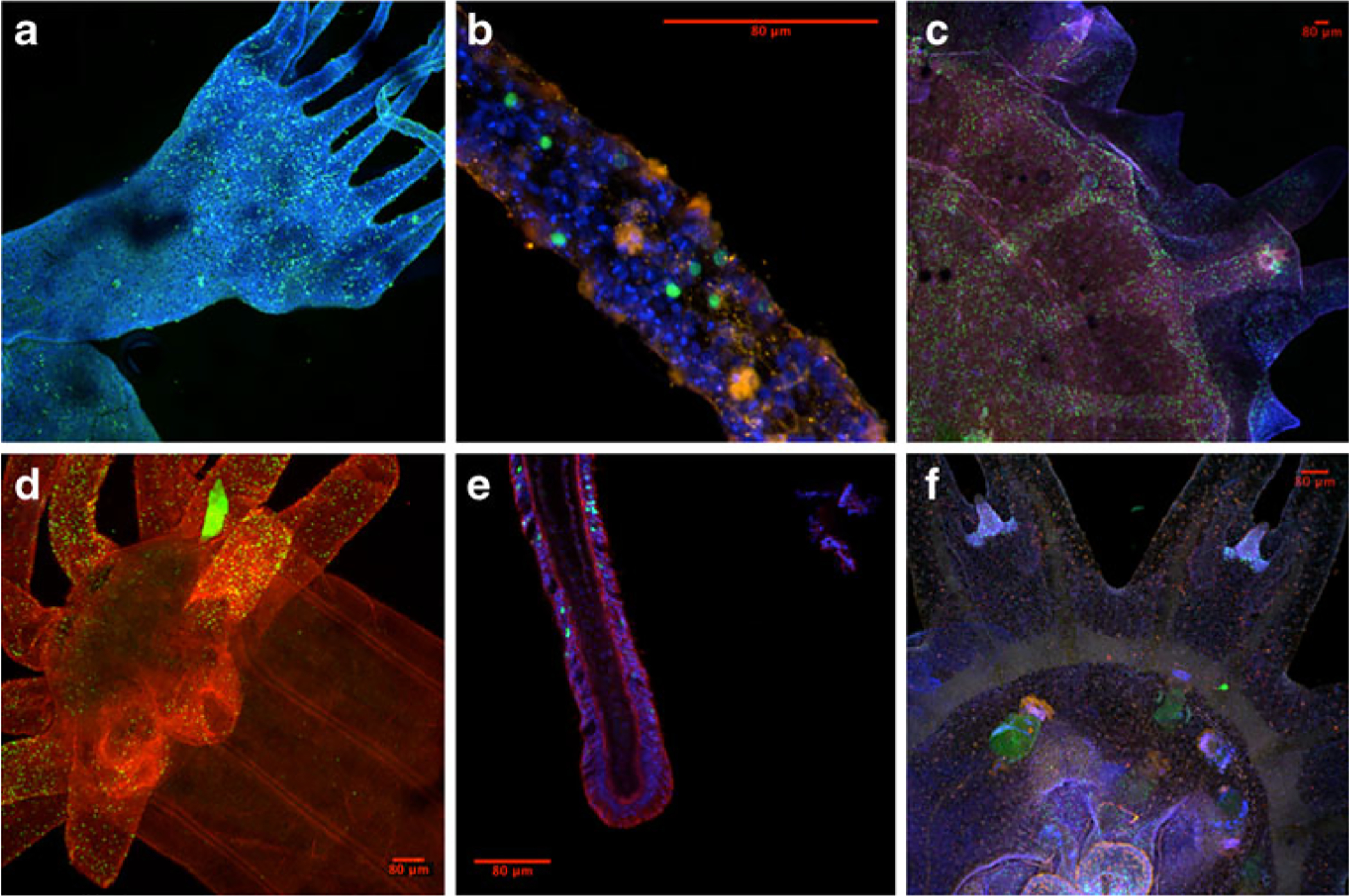
Cell proliferation (in green) in various cnidarians, labeled using 5-ethynyl-2′-deoxyuridine (EdU). a Polyp of Aurelia sp. 1 (nuclear staining in blue); b Close-up of a polyp tentacle (nuclear staining in blue); c Juvenile medusa stage of Aurelia sp. 1 (tyrosinated tubulin in red, nuclear staining in blue); d Polyp of Nematostella vectensis; and e close-up of tentacle (tyrosinated tubulin in red, nuclear staining in blue). f Ephyrae of Chrysaora colorata (fmrf-positive neurons in red, nuclear staining in blue)