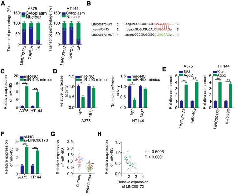
Figure 3.
LINC00173 serves as a ceRNA in melanoma cells and sponges miR-493. (A) Expression localization of LINC00173 in A375 and HT144 cells was identified by a nuclear and cytoplasmic separation assay with RT-qPCR analysis. GAPDH and U6 RNA served as the cytoplasmic and nuclear control transcripts, respectively. (B) The potential miR-493–binding site in LINC00173. Mutant binding sequences are shown too. (C) MiR-493 expression was examined by RT-qPCR analysis in A375 and HT144 cells following transfection with either the miR-493 mimic or miR-NC. (D) Either the miR-493 mimic or miR-NC along with either LINC00173-WT or LINC00173-MUT was introduced into A375 and HT144 cells. The luciferase reporter assay was applied to determine the binding of miR-493 to LINC00173 in melanoma cells. (E) RIP assays were performed to analyze the interaction between miR-493 and LINC00173 in melanoma cells. The enrichment of miR-493 and LINC00173 in A375 and HT144 cells was validated by RT-qPCR. (F) The effects of transfected si-LINC00173 or si-NC on miR-493 expression are shown in A375 and HT144 cells. (G) Total RNA was isolated from the 45 pairs of melanoma tissue samples and adjacent normal tissues and then was subjected to RT-qPCR analysis to evaluate miR-493 expression status. (H) Correlation between miR-493 and LINC00173 expression levels in the 45 melanoma tissue samples was analyzed through Spearman correlation analysis (r = –0.6006, P < 0.0001). *P < 0.05 and **P < 0.01.

