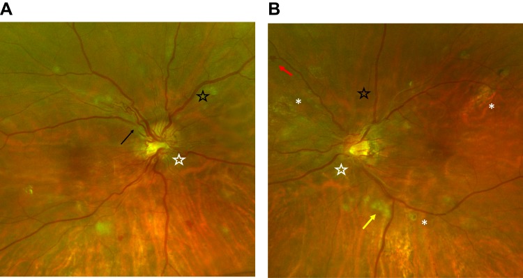
Figure 2.
Optos wide-field color fundus photographs of the right eye (A) and left eye (B) of a patient with hypertensive chorioretinopathy. The photographs demonstrate copper wiring (black star), AV nicking (black arrow), intraretinal hemorrhages (red arrow), cotton wool spots (yellow arrow), Elschnig spots (white asterisk), and optic disc swelling (white star).

