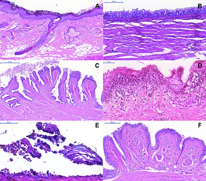
Figure 1.
Histopathological features of dermoid cysts (DCs) (Hematoxylin & Eosin) – (A) Cystic lesion lined by stratified squamous epithelium and hair follicle, sweat gland, sebaceous gland, and adipose tissue in the fibrous capsule (Scale bars = 200 µm). (B) Ciliated pseudostratified columnar (Scale bars = 100 µm) and (C) gut epithelium lining (Scale bars = 500 µm). (D) Chronic inflammatory cells (Scale bars = 50 µm) and (E) multinucleated giant cells (Scale bars = 200 µm). (F) Pacini bodies (Scale bars = 200 µm).

