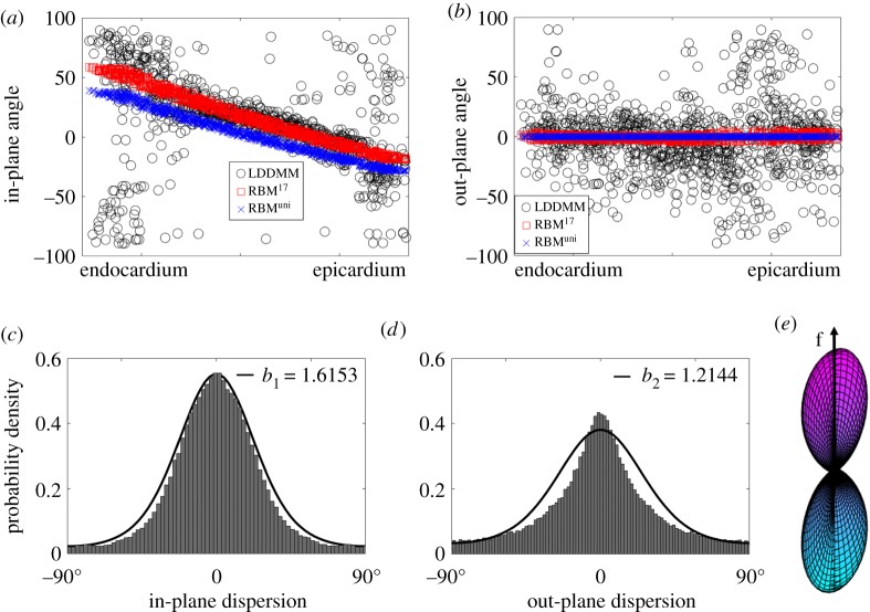
Figure 4.
Fibre dispersion quantified from the DT-MRI dataset. (a) shows the in-plane angle Θ and (b) the out-of-plane angle Φ across the LV ventricular wall; (c) is the in-plane dispersion distribution with fitted ρ(Θ, b1) and (d) is the out-of-plane dispersion distribution with fitted ρ(Φ, b2); (e) a three-dimensional surface plot defined by the vector ρ(Θ, Φ) f(Θ, Φ) with ρ(Θ, Φ) = ρ(Θ, b1) ρ(Φ, b2). The negative angle in (a) suggests the in-plane fibre vector lies in the fourth quadrant (+c0 and −l0), and similarly in (b) for the out-of-plane fibre vector, which lies in the fourth quadrant of plane (−l0 and +r0). All values are used for determining the in-plane and out-of-plane dispersions in (c) and (d).

