Fig. 3.
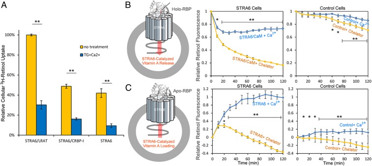
Calcium/calmodulin regulates STRA6-mediated vitamin A transport. (A) Cellular 3H-retinol uptake assay from 3H-retinol/RBP. The same amounts of COS-1 cells were transfected with STRA6/LRAT, STRA6/CRBP-1, or STRA6 alone, and the uptake of 3H-retinol from 3H-retinol/RBP complex was measured by scintillation counting. Thapsigargin (TG), a drug that raises intracellular calcium suppressed cellular retinol uptake. The activity of STRA6/LRAT cells without TG addition is defined as 100%. (B) Real-time retinol fluorescent assay to monitor STRA6-mediated retinol release from holo-RBP. Schematic diagram is shown on the left. 293T cells cotransfected with STRA6 and calmodulin (STRA6 cells) were suspended in HBSS (containing 2 mM calcium) or PBS supplemented with 10 mM EDTA, 20 μM BAPTA [1,2-bis(o-aminophenoxy)ethane-N,N,N′,N′-tetraacetic acid]. Holo-RBP (2 μM) was added at 0 min to start each reaction. Retinol fluorescence was continuously monitored, and its value at 0 min is defined as 1. Lower cellular calcium enhanced STRA6 mediated retinol release from holo-RBP. Control cells are untransfected cells without STRA6. (C) Retinol fluorescent assay to monitor STRA6 mediated retinol loading into apo-RBP. Schematic diagram is shown at Left. Free retinol (2 μM) was added to suspended 293T cells before 2 μM of apo-RBP was added at 0 min to start each reaction. Retinol fluorescence is continuously monitored. The fluorescence value at 0 min is set as 0, and the largest rise of fluorescence signal (STRA6/CaM + Ca2+) is defined as 1. Lower cellular calcium suppressed STRA6-mediated retinol loading into apo-RBP. Control cells are untransfected cells without STRA6. *P < 0.05, **P < 0.05.

