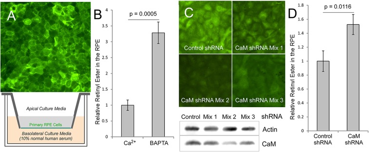
Fig. 4.
Primary RPE cell culture model of vitamin A uptake using human serum as the source of RBP. (A) The primary RPE culture. (Upper) Expression of STRA6 in the RPE as shown by immunofluorescence staining of the primary RPE cells derived from the naturally nonpigmented region of tapetum lucidum. (Lower) Schematic diagram of the transwell culture of primary RPE cells. During each vitamin A uptake experiment, normal human serum, which is a source of holo-RBP, is added to the basolateral culture media to initiate each experiment. (B) HPLC analysis of retinyl ester levels in the RPE after vitamin A uptake from human serum. BAPTA (20 μM), a calcium-specific chelator, leads to increased uptake. Control level (without calcium chelation) is defined as 1. (C) Knockdown of calmodulin in the RPE using a mixture of shRNA for each of the three calmodulin genes. Calmodulin shRNA mixture 2 is most effective in knocking down calmodulin expression. (Upper) Immunostaining of calmodulin in the RPE (calmodulin signal in green and DNA in blue). (Lower) Western blot demonstrating the knockdown of calmodulin. (D) Knocking down calmodulin significantly enhances RPE’s vitamin A uptake from holo-RBP, as revealed by retinyl ester analysis using HPLC. Retiny ester level of control shRNA treated cells is defined as 1. *P < 0.05, **P < 0.05.

