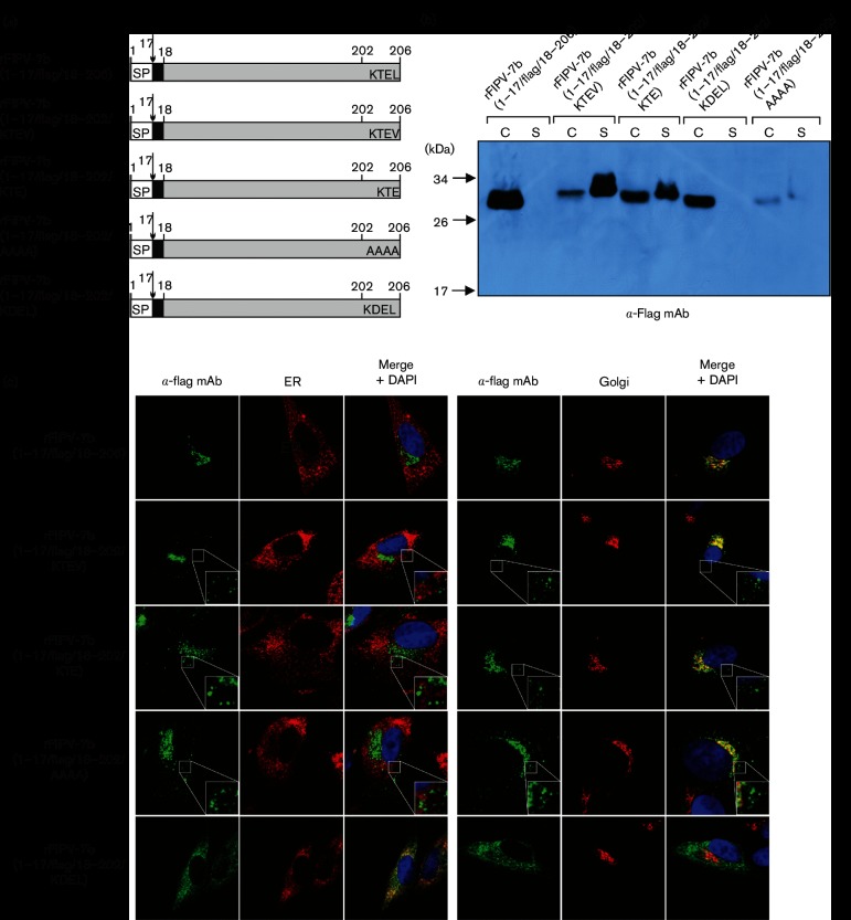
Fig. 4.
The role of the C-terminal tetrapeptide sequence in trafficking and secretion of the FIPV 7b protein. (a) Schematic representation of 7b proteins encoded by rFIPV-7b(1–17/FLAG/18–206), rFIPV-7b(1–17/FLAG/18–202/KTEV), rFIPV-7b(1–17/FLAG/18–202/KTE), rFIPV-7b(1–17/FLAG/18–202/AAAA) and rFIPV-7b(1–17/FLAG/18–202/KDEL). Black boxes represent the position of the FLAG tag in the respective 7b protein variants. The C-terminal amino acids of 7b are shown in the single-letter code; SP, signal peptide; numbers indicate amino acid positions in 7b. The arrow represents the signalase cleavage site. (b) CRFK cells were infected with rFIPV-7b(1–17/FLAG/18–206), rFIPV-7b(1–17/FLAG/18–202/KTEV), rFIPV-7b(1–17/FLAG/18–202/KTE), rFIPV-7b(1–17/FLAG/18–202/KDEL) and rFIPV-7b(1–17/FLAG/18–202/AAAA), respectively, with an m.o.i. of 0.1. Cells and cell culture supernatants were collected at 16 h p.i. Cell lysates (C) and cell culture supernatants (S) were separated by SDS-PAGE (10 %) under reducing conditions and analysed by Western blotting using anti-FLAG mAb (α-FLAG mAb). (c) CRFK cells were mock-infected or infected with rFIPV-7b(1–17/FLAG/18–206), rFIPV-7b(1–17/FLAG/18–202/KTEV), rFIPV-7b(1–17/FLAG/18–202/KTE), rFIPV-7b(1–17/FLAG/18–202/AAAA) and rFIPV-7b(1–17/FLAG/18–202/KDEL), respectively, with an m.o.i. of 1. Cells were fixed at 16 h p.i. and analysed by confocal laser-scanning microscopy. Immunofluorescence staining of 7b was performed using anti-FLAG mAb (α-FLAG mAb, green signal). Immunofluorescence staining of the endoplasmic reticulum (ER) (left panel, red signal) and the Golgi complex (right panel, red signal) is shown. Cell nuclei were stained with DAPI (blue signal). The inserts in the lower right corner represent an 8× magnification of the selected area.

