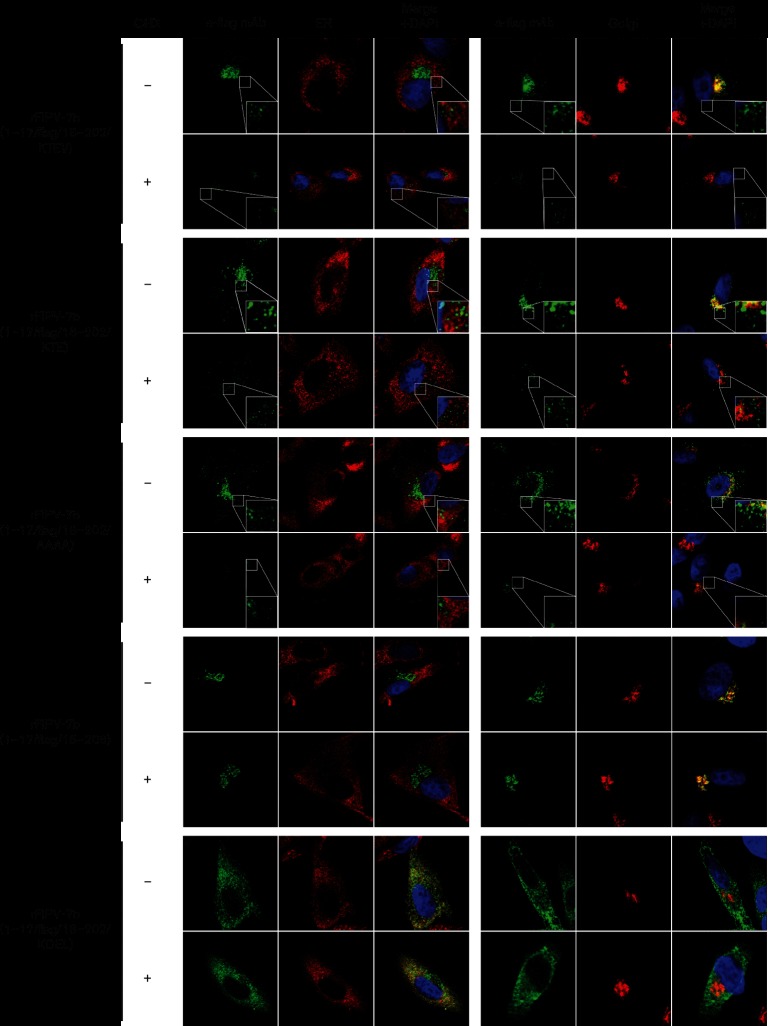
Fig. 5.
Effect of cycloheximide treatment on 7b trafficking. CRFK cells were mock-infected or infected with rFIPV-7b(1–17/FLAG/18–202/KTEV), rFIPV-7b(1–17/FLAG/18–202/KTE), rFIPV-7b(1–17/FLAG/18–202/AAAA), rFIPV-7b(1–17/FLAG/18–206) and rFIPV-7b(1–17/FLAG/18–202/KDEL), respectively, at an m.o.i. of 1. At 15 h p.i., the cells were treated with cycloheximide (+) or left untreated (−). At 17 h p.i., the cells were fixed and analysed by confocal laser-scanning microscopy. Immunofluorescence staining of 7b was performed using anti-FLAG mAb (α-FLAG mAb, green signal). Immunofluorescence staining of the endoplasmic reticulum (ER) (left panel, red signal) and the Golgi complex (right panel, red signal) is shown. Cell nuclei were stained with DAPI (blue signal). CHX, cycloheximide. The inserts in the lower right corner represent an 8× magnification of the selected area.

