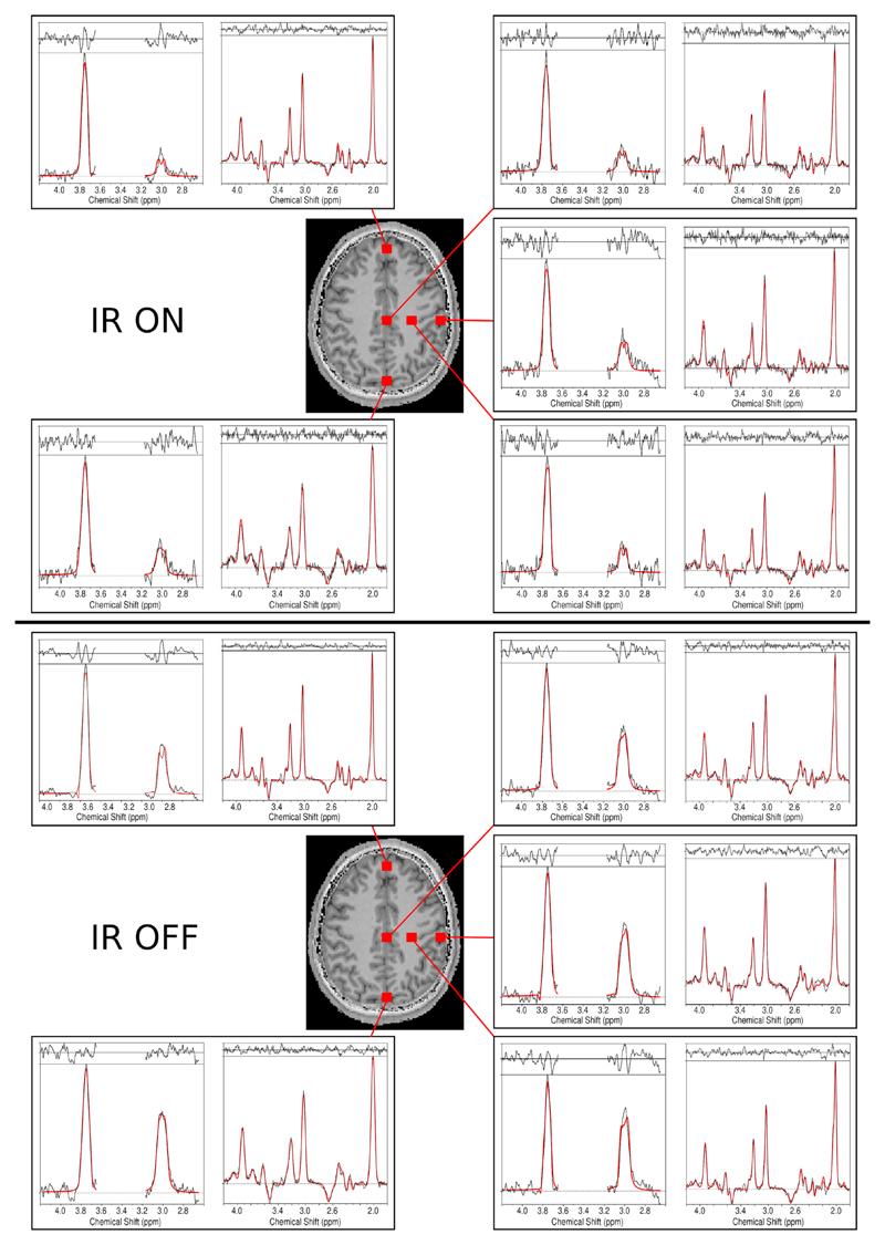
Fig. 5.
Sample spectra including the LCModel fits (in red) for volunteer 1. Spectra from different brain regions that included voxels with typically good spectral quality (mesial gray matter and left frontoparietal white matter) and lower spectral quality (frontal and occipital lobes) are shown. For each voxel, DIFF and EDIT-OFF spectra are grouped together. For comparison, spectra with inversion recovery ON and OFF are provided. A decrease in overall SNR, as well as a significantly reduced GABA peak, is observed in the IR-ON case. The effective voxel size was 1.4 cm3.

