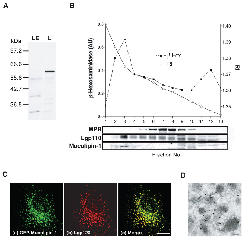
Figure 1. Mucolipin-1 is a lysosomal membrane protein.
(A) Immunoblot of rat mucolipin-1 in rat liver fractions enriched in late endosomes (LE) and lysosomes (L). Proteins (10 μg total per track) were separated by electrophoresis on a 10%SDS polyacrylamide gel using a discontinuous buffer and, after immunoblotting were visualised by ECL. (B) NRK cells were fractionated and the homogenate separated on a 1-22% Ficoll gradient with a 45% Nycodenz cushion. 3 ml fractions were collected from the bottom of the gradient. The refractive index (RI) of the gradient fractions is shown. Each fraction was assayed for β-hexosaminidase activity and immunoblotted for MPR, lgp110 and mucolipin-1. (C) Confocal images (shown as maximum intensity Z projections) using indirect immunofluorescence of (a)GFP-mucolipin-1, (b)lgp120 and (c)extent of co-localisation in NRK cells stably expressing GFP-mucolipin-1. Bar, 10 μm. (D) Immunoelectron micrograph showing GFP-mucolipin-1 (15 nm gold) and lgp110 (10 nm gold) in NRK cells stably expressing GFP-mucolipin-1. Bar, 100 nm.

