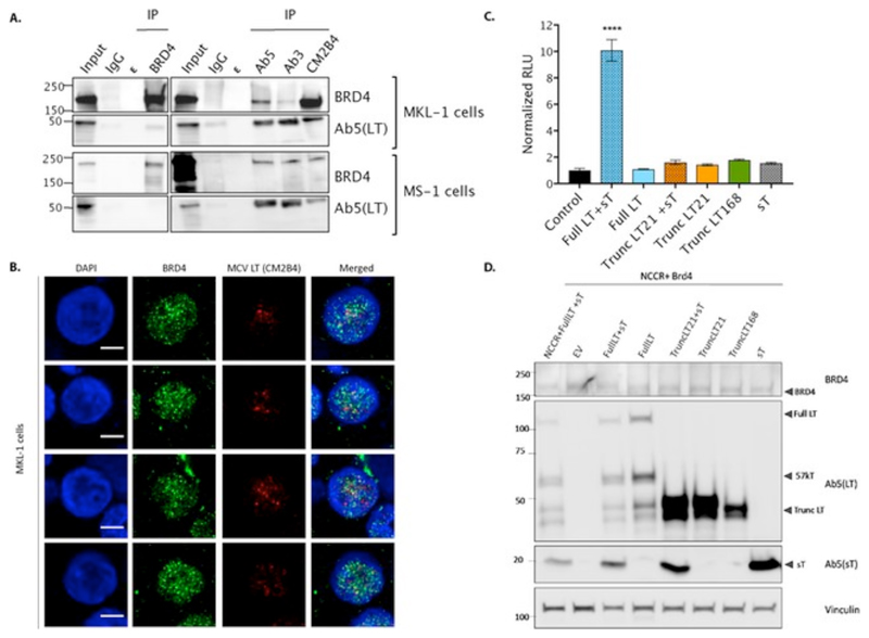
Figure 1. Truncated MCV LT antigen interacts with endogenous BRD4 protein in Merkel cell carcinoma cells.
(A) Nuclear proteins were isolated from MKL-1 and MS-1 cell lines and immunoprecipitated with polyclonal BRD4 antibody and 3 different antibodies against LT antigen i.e. Ab5, Ab3 and CM2B4. BRD4 protein was observed to be co-immunoprecipitated with LT targeting antibodies; however vice-versa was not seen. ε represents the empty lanes between the samples. Input is 2.7% (MKL-1) and 0.8% (MS-1) of total lysate.
(B) MKL-1 were immunostained for BRD4 and LT antigen (using CM2B4) and imaged using FV1000 at 60X magnification. The scale bar represents 5 microns. 4 cells imaged are shown here (of a total of 28 cells, in 3 experiments).
(C) Represents the Relative Luciferase activity in U2OS cells transfected with MCV NCCR region and BRD4 expressing plasmid along with different combinations of MCV T antigen. Two different truncated LT antigens (LT21 and LT168) were used to test increase in luciferase activity. Each column represents the mean value obtained from 3 independent experiments. Error bars represent SD. (2 technical replicates each time). One-way ANOVA with post-hoc Tukey’s test showed Full LT+sT to be statistically significant in comparison to control and other conditions (p<0.0001).
(D) Corresponding western blot for the luciferase analysis confirms the expression of the different T antigen combinations.

