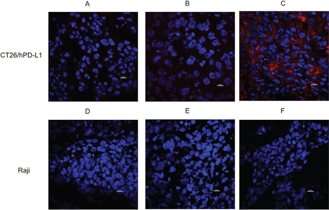
Fig. 5.

Specificity of FN3hPD-L1-01 binder for in vivo detection of hPD-L1 in tumors. (A) Tumor sections from mice bearing CT26/hPD-L1 xenografts were stained with Alexa Fluor 647® 6XHisTag antibody. (B) Tumor sections from mice bearing CT26/hPD-L1 xenografts and injected with 1 mg of FN3hPD-L1-01 binder 24 hours after injection with 100 μg Tecentriq® via tail vein and were stained with Alexa Fluor 647® 6XHisTag antibody. (C) Tumor sections from mice bearing CT26/hPD-L1 xenografts and injected with 1 mg binder via tail vein were stained with Alexa Fluor 647® 6XHisTag antibody (membrane staining). (D) Tumor sections from mice bearing Raji xenografts were stained with Alexa Fluor 647® 6XHisTag antibody. (E) Tumor sections from mice bearing Raji xenografts and injected with 1 mg of FN3hPD-L1-01 binder 24 hours after injection with 100 μg Tecentriq® via tail vein. (F) Tumor sections from mice bearing Raji xenografts and injected with 1 mg binder via tail vein. All sections were stained for the nuclei using DAPI (nuclei staining, center). Image acquisition was performed at 60× magnification using an intravital microscope. Scale bar = 10 μm.
