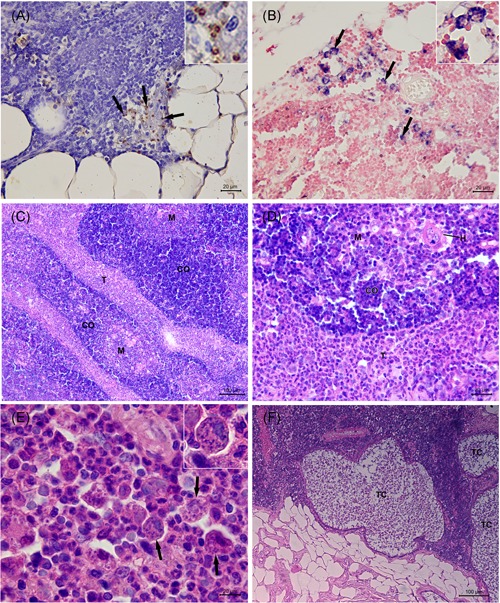
Figure 2.

Histopathological evaluation of the thymus from Leishmania infantum‐naturally infected dogs. A, Brown‐stained amastigote forms of Leishmania spp. (arrow and inset) in the cytoplasm of macrophages (immunohistochemistry). B, Blue‐stained amastigote forms of Leishmania spp. (arrow and inset) in the cytoplasm of macrophages (in situ hybridization). C, Thymitis showing thickening of the interlobular trabeculae due to fibrosis and to an intense pyogranulomatous inflammatory infiltrate (HE). D, Detail of (C) showing an intense pyogranulomatous inflammatory infiltrate in the interlobular trabeculae. E, Detail of (D) showing an intense and poorly formed pyogranulomatous inflammatory infiltrate in the interlobular trabeculae composed mainly by macrophages and neutrophils with few lymphocytes and plasma cells. Several amastigote forms of Leishmania spp. (arrow and inset) are observed within the cytoplasm of macrophages (HE). F, Thymic cysts lined by ciliated epithelium in the parenchyma (HE). C, cortex; H , Hassall's corpuscle; HE, hematoxylin and eosin; M, medulla, T, interlobular trabecula; TC, thymic cyst
