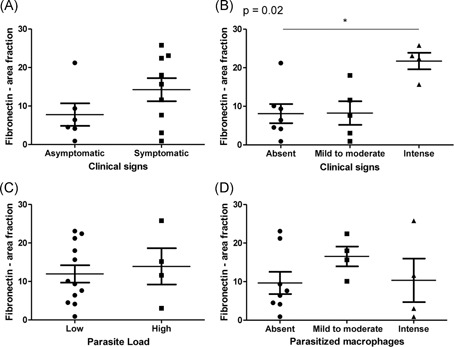
Figure 5.

Analysis of fibronectin deposition in the thymus from L. infantum‐naturally infected dogs. A,B, Comparison of fibronectin deposition between groups according to clinical signs. C,D, Comparison of fibronectin deposition between groups according to parasite load
