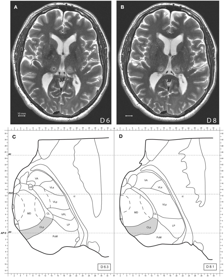
Figure 1.
(A,B) Show axial MR T2 images two days after the treatment, 6 and 8 mm dorsal to the intercommissural plane of a bilateral MRgFUS CLT. (C,D) Show modified atlas maps of the Morel's Atlas 6.3 and 8.1 mm dorsal to the intercommissural plane with the posterior Central Lateral nucleus (CLp) in gray. Mammillothalamic tract (mtt), ventral anterior nucleus (VA), ventral lateral anterior nucleus (VLa), ventral lateral posterior nucleus (VLp), ventral posterior lateral nucleus (VPL), lateral posterior nucleus (LP), medial pulvinar (PuM), mediodorsal nucleus (MD), internal capsule (ic), posterior commissure (pc), anterior commissure (ac).

