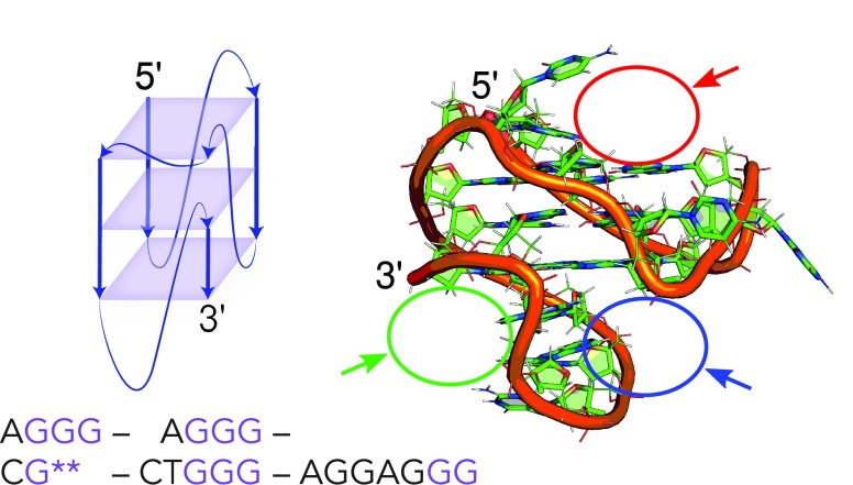
Figure 4.
Schematic representation and structure of ckit-1 showing the binding sites used for docking; G** represents the incomplete tetrad corners filled by Gs at the 3′ end. Red site (1): the 5′-tetrad is openly accessible with potential interaction partners corresponding to C11 and T12. The final AGGAG loop (nucleotides 16 to 20) in ckit-1 folds back to complete the middle and 3′ tetrad, presenting binding sites with the 3′-tetrad (blue site, 2) and without the tetrad (green site, 3).

