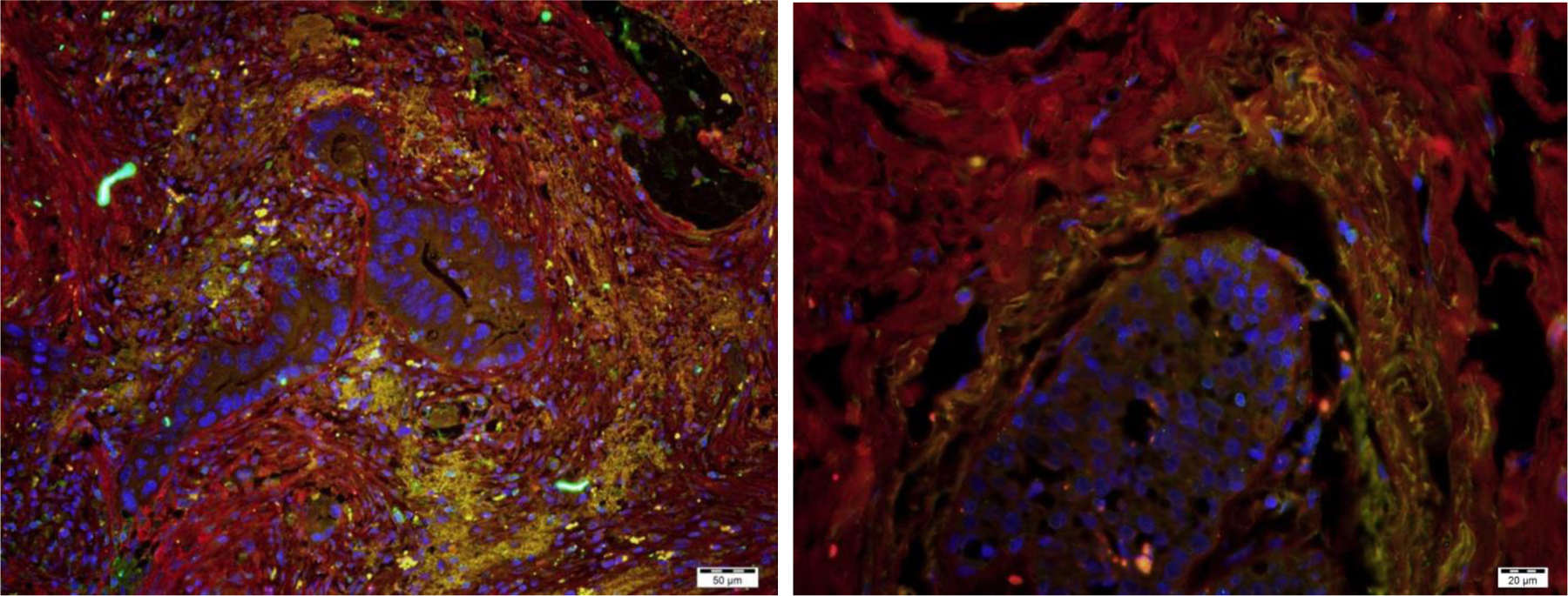
Figure 1:

Fibrocytes surrounding neoplastic ducts in pancreatic adenocarcinoma (left) and invasive ductal carcinoma of the breast (right). The fibrocytes are identified by co-expression (yellow) of Collagen-I (red) and CD45RO (green). DAPI staining of nuclei is shown in blue. Bar is 50 μm on left, 20 μm on right.
