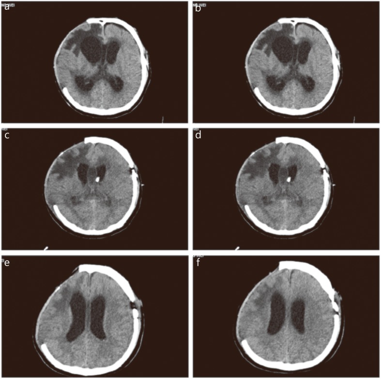
Fig. 2.

Brain CT after admission in patient with intracranial A. baumanni infection. a. Before the treatment on July 3, large low-density shadows were seen in the right frontotemporal parietal lobe; b. Before the treatment on July 3, large low-density shadows appeared in the bilateral lateral ventricles and the third ventricle dilated; c. During the treatment on July 16, bilateral lateral ventricle and the third ventricle were dilated, and hydrocephalus was better than before; d. During the treatment on July 16, a free tube shadow was seen through the left frontal bone to the anterior horn of the left ventricle; e. After treatment on August 2, the original free tube shadow was removed; f. After treatment on August 2, bilateral lateral ventricular hydrocephalus was observed
