Fig. 9.
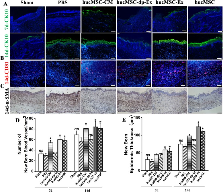
HucMSC-Ex suppresses scar formation and enhances angiogenesis in a full-thickness skin wound model. a Wound histology after immune fluorescence staining. Tissue sections obtained from the wound area at days 7 and 14 after various injections were stained with antibodies against cytokeratin 10 (green). Scale bar = 200 μm. b Tissue sections obtained from the wound area at day 14 after various injections were stained with antibodies against CD31 (red). Scale bar = 200 μm. c α-SMA staining of wound sections treated with hucMSC-CM, hucMSC-dp-Ex, hucMSC-Ex, or hucMSC at day 14. Scale bar = 200 μm. d Quantitative analysis of the number of blood vessels in b. n = 3 per group. *P < 0.05 compared with the PBS group. e Quantitative analysis of the thickness of the new epidermis in a. n = 3 per group. *P < 0.05 compared with the PBS group

