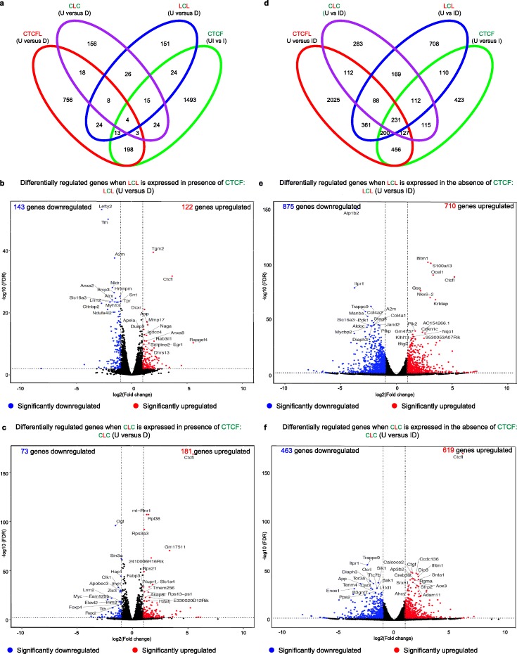
Fig. 5.
Gene expression changes of fusion proteins do not phenocopy that of either parent protein. a, d Venn diagrams showing comparison of deregulated gene expression by CTCFL, LCL, and CLC in the presence (a) and absence (d) of endogenous CTCF. b, c, e, f Volcano plot representation of differentially expressed genes in untreated (U) versus LCL (b, e) and CLC (c, f) expressing mESCs in the presence (D) (b, c) and absence (ID) (e, f) of endogenous CTCF. Red and blue mark the genes with significantly increased or decreased expression, respectively (FDR < 0.01). The x-axis shows the log2 fold-changes in expression and the y-axis the log 10 (false discovery rate) of a gene being differentially expressed. The number of genes that are significantly up- or downregulated is indicated in either case

